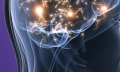
Investigating SUDEP through brain tissue
Understanding the complexities of the brain is key to understanding the causes of epilepsy and the impact of seizures on the brain. It is also pivotal in improving the diagnosis and treatment of epilepsy.
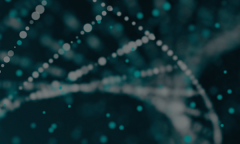
Neuroimaging allows us to look deep inside the brain to map both its structure and, in some cases, to locate the focal point of seizures. Whole genome sequencing is helping us unravel the genetic contribution to epilepsy and how our DNA may be crucial in determining our susceptibility to seizures.
But neuroimaging and genomics still only tell us part of the story. There is still much that we do not know and that is where studies using brain tissue are critical to advance our knowledge of epilepsy at a cellular level.
Dr Joan Liu gives an insight into Epilepsy Society's Brain and Tissue Bank at UCL:
Examining brain tissue is one of the most important ways to help us understand the causes of epilepsy and the impact of seizures on the brain. Which is why we are so grateful to all those who consent to donate their brains, at the end of their lives, to our Epilepsy Society Brain and Tissue Bank.
The brain and tissue bank was established in 2013 and now has almost 400 brains donated by those who have died, and more than 500 surgical samples. These are where a person undergoing brain surgery consents to donate to us any tissue that is removed during surgery. We also have a further 500 brains donated to us from the ERUK Corsellis epilepsy collection.
The epilepsy brain and tissue bank has supported projects in the last five years investigating mechanisms that could underly sudden and unexpected death in epilepsy.
The research has focussed on brain centres known to regulate autonomic respiratory and cardiac functions. If cellular adaptive alterations occur in these regions, because of previous seizures, this may have an effect on modulating the normal cardio-regulatory functions of these centres which could be critical during a further seizure. Alterations in serotonergic and neuropeptide neurotransmitter systems in the brainstem and increased microglial cells in cortical autonomic regions and markers of neuronal activity have been shown and published in research papers (see attached). More research in this area, to explore what could be key cellular alterations, could lead to preventative strategies. We are currently involved in a project exploring the amygdala brain region in epilepsy patients who have had cardiac monitoring during life, to see if there is any direct relationship between similar cellular alterations and autonomic regulatory functions.
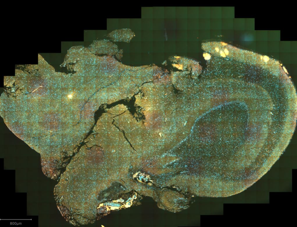
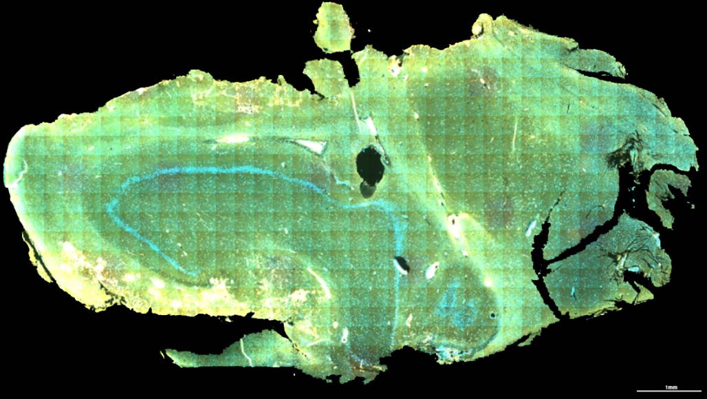
An advanced technique (RNAscope), is able to show gene expression at a cellular level, in this hippocampal tissue kindly donated by people who had undergone surgery as treatment for their Epilepsy.
Understanding SUDEP
One of our key areas of research is to investigate the mechanisms responsible for Sudden Unexpected Death in Epilepsy (SUDEP). This is a leading cause of premature death in epilepsy.
By studying brain tissue samples we have been able to establish a potential link between SUDEP and a depression of the central respiratory system following a seizure, and a link between serotonin pathways and the risk of SUDEP.
Serotonin transmits messages between nerve cells and is also thought to play a role in functions such as heart and respiratory rates.
Brain inflammation and SUDEP
We have also studied neuroinflammation in SUDEP. Brain inflammation is thought to be significant in the excitability of neurones and the progress of epilepsy in the brain.
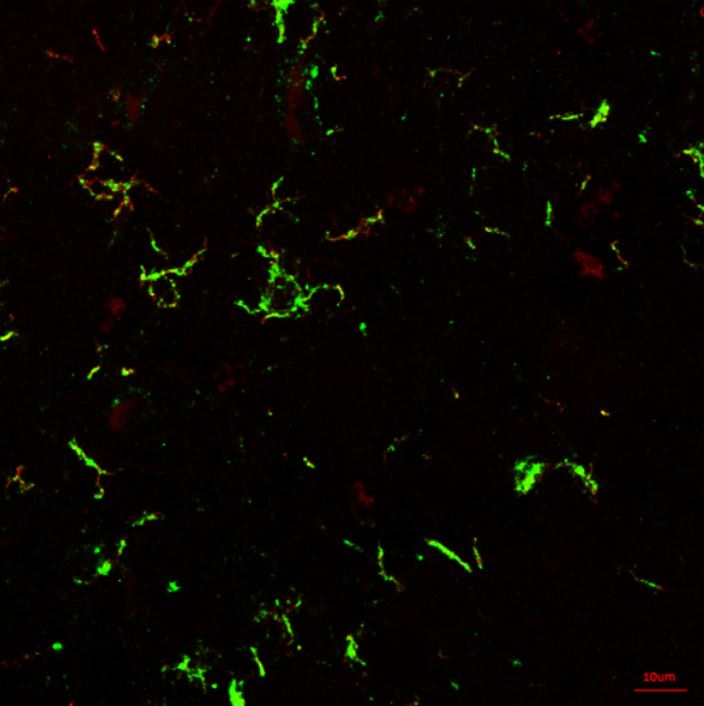
In a pilot study of 55 post mortem samples, we were able to see a significant increase in neuroinflammation in cases involving SUDEP, compared to those with epilepsy but without SUDEP and to disease-free controls.
We also saw an increase in microglia - a type of glial cell that supports and insulates neurones - in areas of the brain that unconsciously regulate bodily functions such as cardiovascular and respiratory responses during a seizure. This raises the possibility of a further link to the cause of sudden death.
Sequencing brain tissue
Our brain tissue samples are preserved in one of two ways. They are either fixed in formalin or they are frozen.
Brain tissue samples donated to us following a person's death, are most commonly fixed. While these are suitable for studying the microanatomy of cells and tissues, it is not so easy to sequence their DNA or RNA (the part of a cell that converts genetic information of DNA into proteins).
Some surgical samples may also be fixed, but many are frozen. This means that we are also able to analyse both the DNA and RNA of the tissue and this is very valuable for our research.
However new technologies are now available that will allow us to analyse the DNA and RNA of fixed tissue samples, offering the potential to enrich our knowledge of the process by which epilepsy develops within a brain.
One of these platforms is called 'Nanostring' and is available through our academic partners, UCL. Now we want to extract DNA and RNA from fixed tissue samples and see if their quality will allow us to use Nanostring technology to analyse information about gene expression.
We hope to then correlate this with information gained through neuropathology, imaging and genomics. This will help us to build up a more comprehensive picture of the causes and biomarkers for SUDEP. If we can identify those at greatest risk, we can also put in place a strategy to manage that risk and ultimately save lives.
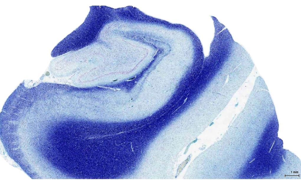
Neuropathology
The Epilepsy Society Brain and Tissue Bank is the first of its kind in the UK. It is dedicated to the study of epilepsy through brain and other tissue samples.
Genomics
Read how we are working to understand the genetic architecture of each individual person's epilepsy through our world leading genomics research programme.
Neuroimaging
Neuroimaging enables us to look deep inside the brain to learn more about the impact of seizures on its structure and function.